Equipment
Zeiss Discovery V12 Stereomicroscope
The Discovery V12 Stereomicroscope is a configurable, modular system featuring newly patented optics for improved resolution and contrast. The motorized zoom system provides resolution, magnification and object field data via a Human Interface Panel (HIP), which is illuminated for easy viewing. The HIP allows programmable entry of eyepiece and objective magnification, zoom speed, focus speed, position and displays resolution, field of view and magnification values. A 3-position turret can accommodate from 0.63X to 1.5X objectives for a zoom range of 30:1.The Microscope is also equipped with a 1K frames per second high speed camera as well as integrated software for CCD image grabbing. Images can be post processed with a wide range of image manipulating software.
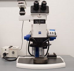
Zeiss Axios Scope
The Zeiss Axio Scope.A1 is a configurable optical microscope offering a range of operation modes including transmission, reflection, polarized microscopy, and both phase and differential interference contrast (DIC) imaging. The microscope is equipped with a binocular phototube, an Axiocam 105 color camera, a removable polarizer, a Nomarski filter slider, a Linkam temperature controlled stage, and a mechanical stage. Available objective lenses include EPIPLAN 5x/0.13 HD, 10x/0.25 HD, 20x/0.4 HD, 50x/0.75 HD, and a 50x/0.55 Neofluar lens.
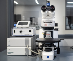
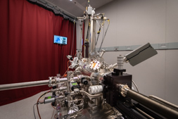
Nicolet iN10 MX
The Thermo Scientific Nicolet iN10 MX is an infrared imaging microscope that offers the ability to rapidly acquire microscopic images and simultaneously collect infrared spectra of solid specimens. It allows analysis of the distribution of chemicals within a sample through both point based analysis and comprehensive spectral mapping. The microscope is equipped with a cooled MCT detector which facilitates ultra-fast imaging and spectral mapping of large areas within minutes. Additional attachments include a MicroTip ATR for contact sampling, nitrogen flow chamber, and a motorized stage. This instrument is ideal for the analysis of coatings, contaminant identification, and the mapping of chemicals in printed media.
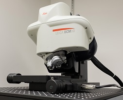
Leica DCM 3D Micro-optical System
The Leica DCM 3D is a dual head confocal and interferometry 3D light microscope. It provides standard bright field microscope imaging, confocal imaging, confocal profiling, PSI, VSI and high resolution thin film roughness measurement on a single instrument. The system has white light and blue LED (λ=460nm) illumination and collects data with either a black/white high resolution CCD camera or a color camera. Its mounted objective lens kit includes: 5X, 10X, 20X, 50X, 150X magnifications and interferometry. LeicaSCAN software controls image acquisition enabling a maximum resolution of 140nm in the x-y plane and an interpolated 1nm resolution on the z axis. Since confocal imaging samples one focal plane (z-axis) at a time, image stacks can easily generate 3D topography maps. Post-acquisition data analysis utilizes Mountains technology Leica Map software.
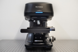
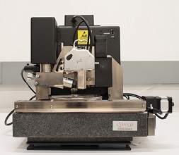
Keyence VK-X3050 Confocal
The Keyence VK-X3050 is a 3D laser scanning confocal microscope. In addition to taking standard bright field microscope images, the laser scanning capabilities allow for non-contact profile measurements used to reconstruct surface topographies and characterize surface roughness and feature heights with nanometer-resolution. Equipped with a red laser (661 nm), the system boasts 5nm resolution in z and 260nm in x-y, and it is capable of resolving sloped surfaces that have angles of up to 78°. The system includes 5X, 10X, 20X, 50X, and 100X (ELWD, WD: 2.0mm) magnification objectives. With a motorized stage, it is also able to stitch together images acquired from multiple fields of view via the stitching and teaching modules.
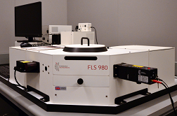
Phosphorescence Fluorescence Lifetime Spectrometer Edinburgh Instruments FLS980
The Edinburgh Instruments FLS980 photoluminescence spectrometer offers both steady state and time resolved (lifetime) fluorescence spectroscopy measurements of solid and liquid samples. The instrument is equipped with four tunable, pulsed repetition rate lasers (wavelengths including: 315, 375, 635, and 980 nm), a flashlamp, and a xenon arc lamp. The FLS980 incorporates two silicon detectors (200-1100 nm) and an InGaAs detector. By combining time correlated single photon counting (TCSPC) and multi-channel scaling (MCS) the FLS980 excels in fluorescence lifetime measurements which span 12 orders of magnitude, ranging from picoseconds to seconds.
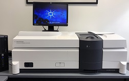
UV-VIS Cary 5000 Spectrometer
The Agilent Technologies Cary 5000 UV-Vis-NIR spectrometer offers high quality photometric performance within a 175 3300 nm spectral range. The Cary 5000 performs absorbance and transmission measurements of both solid and liquid samples. Additional configurations allow the incorporation of an integrating sphere to facilitate external diffuse reflectance measurements. Also, a Universal Measurement Attachment (UMA) is available to perform angle-dependent reflection and transmission measurements.
Verios 460 Extreme High Resolution Scanning Electron Microscope (XHR SEM)
The Verios 460 is capable of sub-nanometer characterization of materials. Super high contrast and image quality can be preserved by integrated beam decelerator, which maintains primary beam coherency, but a low landing energy. Elstar Schottky monochromated (UC) field emission gun (FEG) column provides sub-nanometer resolution (0.6 - 0.7nm) performance from 1 to 30 kV integrated beam deceleration allowing beam landing energies as low as 20 eV piezo-enabled 100x100 mm 5-axis motorized eucentric stage advanced detectors including Everhart-Thornley SED, in-column & below-the-lens detectors, SmartScan and Oxford energy dispersive X-ray spectrometer (EDS) system for elemental mapping and analysis

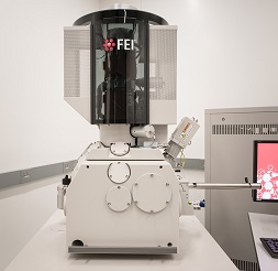
Quanta 200 FEG Environmental-SEM
The Quanta 200 FEG ESEM is a special type of high performance scanning electron microscope (SEM). It is equipped with a Schottky field emission gun (FEG) for optimal spatial resolution. The instrument can be used in high vacuum mode (HV), low-vacuum mode (LV) and the so called ESEM (Environmental SEM) mode. This makes it possible to study samples in pressures up to 30 Torr. The microscope is equipped with a peltier stage for studies of wet samples in-situ. There are cooling and heating stages available for in-situ experiments at temperatures ranging from -20°C up to 1500°C. This can be done in combination with having an external gas present in the chamber, e.g. air, for studying chemical reactions, such as oxidation, at high temperatures.
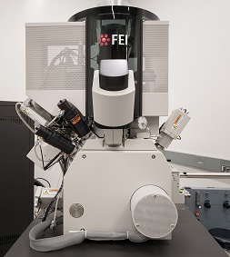
Helios NanoLab G3 UC DualBeam (FIB/SEM)
The FIB/SEM dual-beam system provides the unique capability to add or subtract material at precisely defined locations with high spatial resolution. Its integrated nano-manipulator allows preparation of TEM lamellas. 3D reconstructions is enabled through a "slice and view" before computationally recombining into a single 3D volume. Additional features include:
- An XHR SEM monochromated (UC) field emission electron gun (FEG), constant power lens optics and beam decelerator identical to the Verios for its SEM component.
- Its Tomahawk ion column features superior high current performance (65 nA max beam current) accelerating voltage from 500 - 30 kV, 2-stage differential pumping and time-of-flight correction for a tighter beam and more accurate scan profile.
- Available gas injections are: platinum deposition, carbon deposition, selective carbon mill, and insulator enhanced etch.
- Oxford energy dispersive X-ray spectrometer (EDS) for elemental mapping and analysis.
- EasyLift EX NanoManipulator for easy TEM lamella creation and lift-out.
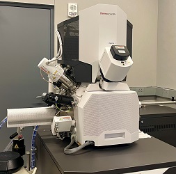
Aquilos 2 Cryo-Focused Ion Beam (Cryo-FIB)
(Available in January 2022)
This is a second-generation dedicated cryo-DualBeam focused ion beam microscope for preparing cryo-lamella samples for TEM tomography of frozen-hydrated biological materials. It is equipped with:
This is a second-generation dedicated cryo-DualBeam focused ion beam microscope for preparing cryo-lamella samples for TEM tomography of frozen-hydrated biological materials. It is equipped with:
- NICol UHR non-immersion field emission-SEM column and Sidewinder ion column.
- In-lens detection system: segmented lower (T1) and upper detector (T2), Everhart-Thornley SE Detector (ETD), ion conversion and electron (ICE) detector for secondary ions and secondary electrons.
- 110 x 110 mm eucentric stage, Fully rotatable cryo-stage (< -170° C at the sample), Tilt range at cryo (eucentric WD): -15° to +55°.
- Automatic Aperture System.
- CCD IR Camera, In-chamber Nav-Cam.
- Cryo Kit, incl. Cryo transfer system, Cryo loading station, conductive deposition GIS, In-chamber retractable sputter coater, EasyLift nanomanipulator, Cryo-FIB AutoGrids.
- Maps 3 software for S/TEM with correlative workflow, AutoScript DB academic, AutoSlice & View, AutoTEM, Cryo for milling automation, SPI™ and iSPI™ software, etc.
- Integrative Fluorescence Light Microscope (iFLM) correlative fluorescence imaging and ion milling
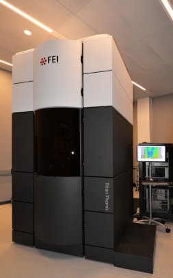
Titan Cubed Themis 300 double Cs-corrected Scanning/Transmission Electron Microscope (S/TEM)
The premiere aberration-corrected S/TEM for atomic-resolution characterization, including differential phase contrast (DPC), bright field, dark field and high-angle annular dark field imaging, electron diffraction, and quantitative elemental analysis/mapping, and EELS acquisition.
- A Schottky X-FEG high brightness electron source with 0.07 nm (TEM), 0.06 nm (STEM) information limit.
- Double Cs-corrector DCOR (Cs image + DCOR probe corrector).
- Flexible high tension range from 60 to 300 kV.
- Symmetric Ruska-Rieke constant power S-TWIN objective lens for thermal stability and easy mode switching.
- Industry-leading high-speed, high-throughput, windowless 4-detector, Super-X energy dispersive X-ray spectroscopy (EDS) signal detection.
- A Gatan Quantum SE/963 P post-column energy filter equipped with a 2k x 2k CCD for energy filtered TEM (EFTEM) and UltraFast (up to 1000sp/s) electron energy-loss spectra (EELS) data acquisition.
- Differential phase contrast (DPC) imaging for electromagnetic studies.
- Automatic apertures and reproducible recall.
- Rotation-free imaging for clear orientation.
- Computerized 5-axis specimen stage with piezo-enabled drift compensation.
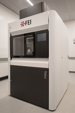
Talos F200X Scanning/Transmission Electron Microscope (S/TEM)
Room temperature S/TEM imaging and analysis including differential phase contrast (DPC), bright field, dark field and high-angle annular dark field imaging, electron diffraction, highly sensitive 4-detector Super-X EDS for quantitative elemental analysis and mapping.
- X-FEG high brightness electron source (1.8x10⁹ A/cm2srad @200 kV) with 0.12 nm (TEM), 0.16 nm (STEM) information limit.
- Flexible high tension range from 60 to 200 kV.
- Constant power A-TWIN objective lens for thermal stability and easy mode switching.
- Industry-leading high-speed, high-throughput, windowless 4-detector, Super-X energy dispersive X-ray spectroscopy (EDS) signal detection.
- Differential phase contrast (DPC) imaging for electromagnetic studies.
- Automatic apertures and reproducible recall.
- Rotation-free imaging for clear orientation.
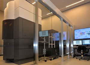
Titan Krios G3 cryo Transmission Electron Microscope (cryo-TEM)
The Titan Krios G3 cryo transmission electron microscope (cryo-TEM) is the most powerful high resolution electron microscope specializing in cryo-based 3D characterization of biological samples. It is equipped with:
- A Schottky X-FEG high brightness electron source.
- Cs image corrector for compensation of spherical aberration (Cs) and semi-automated correction of astigmatism [0.08 nm (TEM) information limit].
- Flexible high tension range from 60 to 300 kV.
- Symmetric constant power C-TWIN objective lens with wide pole piece gap of 11 mm.
- Cryo autoloader for robotic loading of up to 12 frozen, hydrated samples.
- Volta phase plate solution for tunable phase shifting to optimize contrast.
- Low dose software kit to minimize electron dosage.
- Semi-automated data collection for continuous multi-day data acquisition.
- Falcon 3EC pre-GIF direct electron detector camera.
- Selectris energy filter coupled to a Falcon 4i direct electron detector camera..
- Automatic apertures and reproducible recall.
- Rotation-free imaging for clear orientation.
- A computerized 5-axis specimen stage with piezo-enabled drift compensation.

Titan Krios G4 cryo Transmission Electron Microscope (Cryo-TEM)
(Available in January 2022)
This is a new generation of high-resolution electron microscope for high-speed single particles analysis, cellular tomography and micro electron diffraction (MicroED). It is equipped with:
This is a new generation of high-resolution electron microscope for high-speed single particles analysis, cellular tomography and micro electron diffraction (MicroED). It is equipped with:
- A Schottky X-FEG high brightness electron source.
- Flexible high-tension ranges from 60 to 300 kV with high-stability generator.
- A five-lens condenser system with symmetric constant power C-TWIN objective lens, etc. for parallel illumination over a wide and variable field of view.
- High-speed drift correction, Semi-automated data collection for continuous multi-day data acquisition.
- Low dose software kit to minimize electron dosage.
- Selectris energy filter coupled to a Falcon 4i direct electron detector camera.
- Automatic apertures and reproducible recall.
- Rotation-free imaging for clear orientation.
- A computerized 4-axis specimen stage with piezo-enabled drift compensation, and ±70 degree alpha-tilt.
- Cryo autoloader for robotic loading of up to 12 frozen, hydrated samples.
- TEM tomography data acquisition software, Velox imaging software, linear distortion correction, MicroED package, etc.
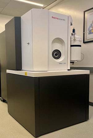
Talos L120C G2 Transmission Electron Microscope
Versatile TEM system designed for modular versatility, maximum stability, and operational ease. Designed for optimized imaging of Pathology, Cell Biology, and soft matter research at ambient and cryogenic (liquid-nitrogen) temperatures. With added automation for tomography and single particle analysis screening.
- Thermionic source (LaB6 emitter) for stable bright images.
- Flexible high-tension ranges from 40 to 120kV.
- Constant-Power C-TWIN lens for thermal stability.
- Quick sample exchange for quick contamination-free imaging recovery.
- Rotation-free imaging for clear orientation of samples.
- 4k x 4k Ceta 16M CMOS camera provides a large field of view with high speed and sensitivity.
- Observe cryo or room temperature samples.
- Automatic C2 and objective aperture and cryo box for reproducible recall.
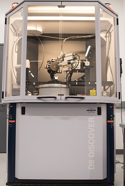
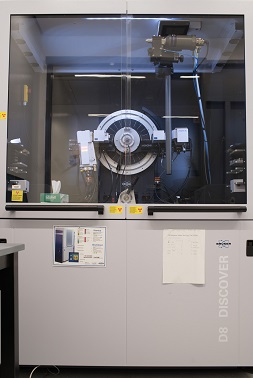
Bruker D8 Discover X-Ray Diffractometer
This XRD is a state-of-the-art, high-quality system that is easy to use, highly accurate, and versatile to meet our needs in X-ray diffraction. This system equips with the optimum components (X-ray 4-Bounce monochromator, high-precision, two-circle goniometer, LynxEye detector and standard software package) to provide a comprehensive solution for X-ray reflectometry, High resolution X-ray diffraction, Grazing incidence diffraction and texture measurements, as well as ?m-XRD. One of the unique advantages of this system is its extreme versatility. The system can be easily reconfigured for the usage of multiple research groups for different applications. The combination of various measurement methods in this single instrument allows us to use it during all phases of complex materials development. We have also added special attachments to meet our research needs. These features include:
- A new LynxEye detector that operates both in a 1-Dimensional Mode (for polymers and powders) and also in a 0 Dimensional Mode (for thin film and grazing incidence analysis, reciprocal space mapping) provides high resolution detection for nearly all applications.
- A Laser-Video Scope for accurate alignment and positioning of any type of samples.
- A DCS 350 temperature stage (-100°C to 350°C).
- A MRI Hot Humidity Stage (25°C - 90°C, up to 90% relative humidity).
- A full TOPAS and MULTEX software package for powder pattern fitting, pattern decomposition, quantitative structure refinement, ab-initio structure determination, and pole figure measurement, etc.
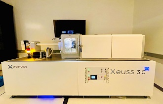
Small-angle X-ray Scattering System (SAXS)
The Xenocs Xeuss 3.0 serves as an in-lab beamline with the ability to deliver both small-angle and wide-angle X-ray scattering capabilities (SAXS and WAXS). SAXS / WAXS involves the elastic scattering of X-rays across small features at angles ranging from ~0.1 to 10 degrees. These techniques enable the assessment of particle ordering and the ability to measure material features which range in size from 1.0 to 300 nm. X-ray scattering is useful in the analysis of colloids, surfactants, mesopores, block copolymers, and biological structures. The IACs SAXS harnesses a Genix 3D X-ray source with a dynamic range of sample-to-detector distances extending from 4.5 to 105 cm and the offers the ability to perform grazing incidence (GISAXS). Additional holders enable the analysis of powders, liquids, and gels at temperatures ranging from -150 to 350 degrees C. XSACT software enables users to rapidly produce publication quality data.
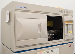
Rigaku MiniFlex XRD
Rigaku MiniFlex XRD/X-ray diffractometer equipped with two types of holders for powder and bulk) to have up to 6 samples which can be mounted and measured with the automatic sample changer. Each sample can be made to spin to enhance data precision for the qualitative and quantitative analyses. It works with JADETM 3.1 processing and search/match software.
Bruker D8 Advance X-Ray Diffractometer
The Bruker D8 Advance X-ray diffractometer (XRD) is a dedicated powder XRD designed to extract structural information from standard or small volume samples. Primary, secondary optics, detectors and sample stage are modular in design and swappable to fit experimental needs. The DAVINCI module of the operating software smartly recognizes the installed components and adapts the data collection parameters to accommodate. In addition to standard Bragg-Brentano geometry, the system can also perform grazing incidence and small-angle X-ray scattering (SAXS) measurements.
Features include:
Features include:
- X-ray source (Cu or Ag) flexibility for different samples.
- LynxEye XE high-resolution energy-dispersive detector for 0D, 1D and 2D diffraction.
- Flipstick 9-position sample changer for automatic loading and data collection of multiple samples.
- Capillary transmission sample mounting for small volume samples.
- HTK 1200N temperature chamber for in-situ characterization (25 - 1200°C).
- Oxford cryostream 700 series with cold stream deflector for integrated LN2 enabled capillary sample cooling.
- Diffrac.EVA software for data analysis.
- Diffrac.NANOFIT software for SAXS analysis.

Zeiss Xradia 630 Versa High-resolution 3D X-ray tomography Microscope
The Zeiss Xradia 630 Versa is a non-destructive submicron 3D X-ray microscope. Its industry-leading resolution and contrast achieves spatial resolution of 0.7 μm and minimal achievable voxel of 70 nm. This system bridges the resolution gap between light and electron microscopes enabling easy, non-destructive 3D reconstructions of samples. It is equipped with:
- Dual Scan Contrast Visualizer (DSCoVer), enabling compositional probing through overlay of imaging data of a single sample at two different X-ray spectra.
- High-Aspect Ratio Tomography (HART) for higher throughput imaging for flat samples like semiconductor packages and boards.
- Automated filter changer (AFC) for easy X-ray spectrum tuning.
- Wide field mode & vertical stitching for extended imaging of larger samples.
- Flat panel extension (FPX) to image significantly larger samples (beyond 5" diameter).
- CT50000-TEC in-situ interface kit for heating, cooling, tension and compression stage.
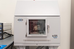
Bruker Dimension ICON3 Atomic Force Microscope
The Icon3® atomic force microscope (AFM) introduces new levels of performance, functionality, and AFM accessibility to nanoscale researchers in science and industry; high performance, multifunctional, scanning probe microscope (SPM) optimized for peak force characterization.
- Housed within an integrated acoustic and vibration isolation enclosure to ensure optimal data collection.
- Motorized stage with a typical X-Y scan range of 90μm x 90μm.
- Various advanced operation modes including: PeakForce Tapping/contacting, Force spectroscopy, Surface potential, Piezoresponse microscopy, Tunneling AFM, etc.
CreaTec LT STM/nc-AFM
The CreaTec Special Low-Temperature STM type LT-STM-AFM-CE system rely on the combination of Scanning Tunneling Microscopy (STM) with qPlus based Atomic Force Microscopy (AFM) techniques. While STM can reveal band structures, electronic states for materials, electron orbital related signal for molecules, qPlus based non-contact AFM offers sub-molecular real-space atomic resolution for materials and organic molecules. This system is mainly used to study the electronic states, electron orbitals and the atomic structure information of molecules, to identify the chemical structures of unknown organic compounds. Low temperature (4K) constant current STM imaging, scanning tunneling spectroscopy (STS), constant frequency AFM imaging, constant height AFM imaging, and force spectroscopy measurement can be conducted.
Bruker NanoMan AFM
The NanoMan AFM incorporates the Dimension platform, the advanced NanoScope V controller and the Hybrid XYZ scanner to create the preeminent system for high-resolution imaging, high-definition nanolithography, and direct nanoscale manipulation. The NanoMan is also able to perform highly accurate force curves, nanoindenting and pulling techniques. This state-of-the-art AFM operates in closed-loop mode, enabling a single instrument solution for nanolithography, nanomanipulation, and imaging for material and life science applications.
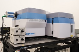
Horiba Raman Spectrometer
Raman spectroscopy is a vibrational spectroscopy that uses light scattering to infer vibrational spectra of solid samples. It can help in identification of molecular compounds, bonding geometries, chemical mapping, etc. It can be used for applications as wide ranging as pharmaceuticals, forensic science, polymers, thin films, semiconductors and even for the analysis of fullerene structures and carbon nano-materials. Our Horiba Aramis spectrometer has confocal and micro-mapping capabilities, and it offers a selection of 325, 532, 633, and 785 nm excitation lasers.

Thermo Nexsa G2 X-ray Photoelectron Spectrometer (XPS/UPS/ISS/Raman)
ThermoFisher Nexsa G2 is the next generation X-ray Photoelectron Spectrometer (XPS) that gives quantitative elemental composition and chemical state information about the sample surface (top 3 10 nm). Additional tools allow to perform ion etching, depth profiling, UPS, Raman, and ISS spectroscopies. The system contains:
- Al Kα monochromated, focused X-ray source with variable spot size (10 400 µm)
- Ar+ ion gun with ion energy range of 0.3 4 keV and monoatomic/cluster modes
- Ultraviolet Photoelectron Spectroscopy (UPS) with He I/He II UV light source
- Raman spectroscopy with a 532 nm laser
- Ion Scattering Spectroscopy (ISS) with He ion source
- Dual electron/ion flood gun for charge compensation
- Sample holders include: vacuum transfer module, heating stage, SEM integration unit
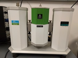
Woollam M-2000 Ellipsometer
The M-2000® spectroscopic ellipsometer is engineered to meet the diverse demands of thin film characterization. An advanced optical design allows to determine the thickness and the optical parameters (index of refraction and absorption coefficient) of films from few nanometers up to few microns thick. The instrument covers 190 1700 nm spectral range. Mapping mode allows to visualize film thickness variations across the sample. Temperature controlled sample stage is available to observe thermal dynamics of films in the -50 to 350 °C range.
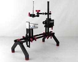
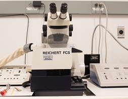
RAMÉ-HART Goniometer
The ramé-hart Model Goniometer is a tool for contact angle measurement. Model 90 includes DROPimage software as well as our German-made SuperSpeed U3 Series digital camera with 1920 x 1080 resolution, LED backlight, leveling specimen stage, and highly modular and flexible optical bench. The measurements can be performed on droplets on the surfaces and on droplets suspended from a needle. With DROPimage Pro software the instrument is capable of measuring contact angle, surface energy, surface tension, and interfacial tension.

Thermo K-Alpha+ X-ray Photoelectron Spectrometer (XPS/UPS)
ThermoFisher K-Alpha+ is an X-Ray Photoelectron Spectrometer (XPS) that gives quantitative elemental composition and chemical state information about the surface (top 3 10 nm). Additional tools allow to perform ion etching, depth profiling, and UPS spectroscopy. The system contains:
- Al Kα monochromated, focused X-ray source with variable spot size (30 400 µm)
- Ar+ ion gun with ion energy range of 0.3 4 keV and monoatomic/cluster modes
- Ultraviolet Photoelectron Spectroscopy (UPS) with He I/He II UV light source
- Electron flood gun for charge compensation
- Sample holders include vacuum transfer module

PerkinElmer DSC-8500 Differential Scanning Calorimeter (DSC)
The DSC-8500 is a double-furnace automated multi-sample high performance differential scanning calorimeter. It features a second generation high speed DSC (HyperDSC) and is capable of: extremely fast scanning rates to 750°C/min, in-situ ballistic cooling 2100°C/min, with fast data readout rates (100 points/second) providing high data integrity. This allows for quick and accurate thermal characterization of heat capacity and phase transitions.
PerkinElmer DMA-8000
Dynamic Mechanical Analyzer (DMA)
The DMA-8000 is a temperature-controlled dynamic mechanical analyzer. It features a 180° rotating analysis head enabling configuration for any test type and sample geometry. It couples a LN2 dewar with a standard furnace for temperature control between -190°C to 400°C. The system is capable of dynamic oscillations ranging from 0 to 600 Hz. And in addition to operation in dynamic mode, also features "Constant force (TMA) mode" vs. time or temperature. Combined, this system is capable of determining a material's dynamic modulus (storage, tan(δ)), stress/strain, viscoelastic properties, and even expansion coefficient (softening/penetration).
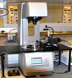
Anton Paar MCR702 Rheometer
The Anton Paar MCR 702 is a two motors rheometer capable to operate both in strain and stress controlled mode. It is equipped with the classical measuring geometries spanning smooth and rough surfaces, parallel-plates, cone-plate, large and small diameters. Mechanical and flow properties such as elastic modulus, viscous modulus, viscosity and normal stresses as a function of stress or strain, of a broad class of materials ranging from water-like fluids, polymers to cement pastes can be measured. The instrument is also capable of Rheo-Optical measurements by a microscope coupled with the rheometer thus allowing rheology with optical characterization of microstructure.
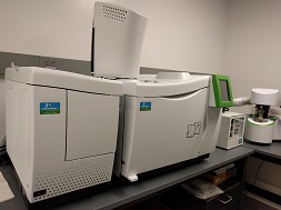
PerkinElmer TGA-GC/MS: TGA-8000
Thermogravimetric Analyzer (TGA),
Clarus 680 Gas Chromatography (GC),
Clarus SQ 8 T Mass Spectrometry (MS) hyphenated system
Three instruments combined into one flexible workflow, the TGA-GC/MS enables use of one or more instruments in thermal characterization. TGA can be used alone to analyze the mass loss or gain due to temperature-driven decomposition, oxidation or loss of volatiles. Through a high accuracy measurement of mass change, temperature and temperature change, both physical and chemical phenomena can be observed. This allows for analysis of second-order phase transitions such as vaporization, sublimation, absorption, adsorption, desorption along with chemisorptions, desolvation/dehydration, decomposition and solid-gas reactions. When coupled with mass spectrometry via TGA-MS, the TGA-evolved gas product can be fed into the MS for identification. For full use of the hyphenated system, TGA-GC/MS, TGA-evolved gas product can be fed into a gas chromatograph (GC) for further separation before then passing the GC product into the MS for identification/analysis.
Anton Paar MCR501 Rheometer
The Anton Paar Physica Modular Compact Rheometer (MCR-501) applies a controlled stress (torque) to a sample and measures the strain (rotation). As such, the MCR is particularly well-suited to making measurements at very low deformation rates, which are typically required if the intrinsic response of microstructured materials is desired (higher deformation rates disrupt the microstructure). The MCR can access steady shear rates as low as 10-6 sec-1 and as high as 103 sec-1 and conduct dynamic oscillatory measurements as low as 10-4rad/sec; cone-and-plate, parallel plate and Couette geometries are available, and temperature control can be achieved with a circulating water bath, a Peltier plate, or with an electric plate.

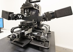
Rheo-Confocal Microscope
The Rheo-Confocal Microscope system combines an Anton Paar MCR502 WESP rheometer with an inverted Nikon Ti-Eclipse confocal microscope equipped with a spinning disk and solid-state laser system. The Rheo-Confocal system allows rheometrical measurements coupled with optical characterization of the microstructure that cannot be achieved with bright field microscope. This system is ideal for visual characterization of bacterial biofilms, intracellular dynamics, and dense particle suspensions while simultaneously conducting rheological measurements of a system.
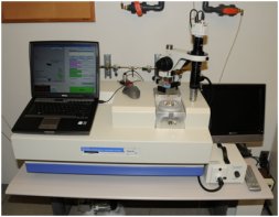
Leica Ultracut UCT Ultramicrotome
The Leica Ultracut UCT ultramicrotome with a cryo-attachment is designed to meet the precise requirements of sample preparation for electron microscopy. It is ideal for biological applications and soft materials preparation. It features automatic feed in 1nm increments from 1 to 100 nm. The variable cutting speed can be finitely controlled to 0.05 mm per second. All key functions of the microtome, even the motorized approach of knife to specimen, can be operated from the separate control unit.

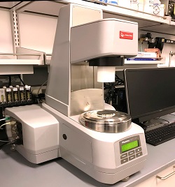
Vitrification Robot
Sample preparation for cryo-electron microscopy by plunge-freezing involves a few steps: application of the sample to a specimen grid, blotting away excess liquid and dropping the thin (about 100 nm) sample into liquid ethane for rapid cooling into a glass-like frozen hydrated state.
The Vitrobot (Vitrification Robot) is a fully PC-controlled device for vitrification (rapid cooling to a glass-like state) of aqueous samples. Environmental parameters relevant to sample vitrification are strictly controllable by dedicated hardware and software, enabling accurate and reproducible results. This level of automation is conducive to high throughput of vitrified samples with easy and straightforward control of the vitrification process through its intuitive user interface. Prior to plunging, loaded samples are maintained inside an environmentally closed enclosure, allowing for control of the following vitrification parameters:
The Vitrobot (Vitrification Robot) is a fully PC-controlled device for vitrification (rapid cooling to a glass-like state) of aqueous samples. Environmental parameters relevant to sample vitrification are strictly controllable by dedicated hardware and software, enabling accurate and reproducible results. This level of automation is conducive to high throughput of vitrified samples with easy and straightforward control of the vitrification process through its intuitive user interface. Prior to plunging, loaded samples are maintained inside an environmentally closed enclosure, allowing for control of the following vitrification parameters:
- Temperature (4 - 60°C).
- Humidity (0 - 100%).
- Blot Time (sec).
- Wait Time (sec).
- Drain Time (sec).
- Blot Force.
- Blot process (# repetitions).

The Leica EM ACE600 Sputter Coater
The Leica EM ACE600 sputter coater offers the rapid ability to simultaneously coat multiple samples in a uniform conductive film. This sample preparation technique is essential in order to achieve optimal resolution during the SEM imaging of non-conductive and beam sensitive samples. Through application of the Leica ACE600, users are capable of depositing either an iridium or carbon coating of size-tunable thickness onto a specimen via plasma sputtering. Full specimen coverage is ensured by utilizing the instruments tilt controls to coat samples at angles ranging from 0 to 45 degrees. The instrument is pre-programmed with standard sputtering sequences which offer rapid film deposition and allows custom programing options for tailored sputterant deposition.
Fischione 1010 Ion Mill
The Model 1010 Ion Mill is a tabletop, PC-controlled precision milling and polishing system for creating high-quality TEM specimens with large electron transparent areas. It is fully programmable and easy to use. The Model 1010 incorporates two independently adjustable, variable energy hollow anode discharge (HAD) ion sources, liquid nitrogen specimen cooling, 0° to 45° milling angles, automatic gas control, and an oil-free vacuum system for ultra-clean specimen processing.
Fischione NanoClean Plasma Cleaner
The NanoClean is an RF Plasma sample cleaning tool that utilizes reactive gas compounds formed by the plasma to chemically reacting with the carbonaceous debris that exists on the sample surface. The gas mixture fed to the plasma contains 21% of oxygen and 79% of argon. The chamber is pumped by oil-free pumping system that is composed of a multistage diaphragm pump and a turbomolecular drag pump. The NanoClean can be used to remove surface carbonaceous contamination from both TEM and bulk samples, and the unit at IAC also comes with one vacuum pumping station that accommodates vacuum storage for up to 5 TEM holders.
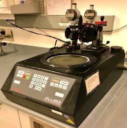
Allied Multi Prep Polisher
The MultiPrep™ System enables precise semi-automatic sample preparation of a wide range of materials for microscopic (optical, SEM, TEM, AFM, etc.) evaluation. Capabilities include parallel polishing, precise angle polishing, site-specific polishing or any combination thereof. It provides reproducible sample results by eliminating inconsistencies between users, regardless of their skill. The MultiPrep eliminates the need for hand-held polishing jigs, and ensures that only the sample makes contact with the abrasive. It maintains geometric orientation of the sample relative to the abrasive plane during polishing, allowing quantification of material removal; rate of polish can be monitored, and total amount removed can be preset.

The VCR IBS/TM200S Ion Beam Sputterer
The VCR IBS/TM200S ion beam sputterer is designed to deposit ultra-fine grain conductive films of metal or carbon on the surface of a sample. The IBS can facilitate the deposition films composed of a variety of materials including iridium, carbon, chromium, platinum, and gold. These films are necessary to reveal the microscopic details made visible by the optimal resolving power of the latest generation of high-resolution scanning electron microscopes (SEMs). The IBS/TM200S deposits films by striking a target material with a plasma beam and sputtering material from the target onto the specimen. This process allows the low-energy deposition of sputterant in order to produce ultra-thin amorphous films, without requiring bombardment of the specimen of interest with high-energy plasma. Additionally, the specimen can be moved in a complex fashion during deposition to ensure uniform film deposition by using independent stage rotation and tilting controls.
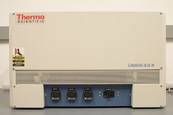
The Thermo Scientific Lindberg Tube Furnace (available soon)
The Thermo Scientific Lindberg Blue M tube furnace offers precise heating of samples under a controlled atmosphere at temperatures ranging from 100 to 1,100 °C. The Lindberg tube furnace is equipped with a 3 in. diameter insertable quartz tube which can be sealed with flanged fittings to allow heating of multiple samples under flowing gas. The furnace employs three programmable PID controllers, each of which control an individual heating zone (i.e. 6, 12, and 6 respective heating zones) to ensure adequate heating is achieved along the length of the tube without overshooting the targeted temperature. The controllers can be programmed to run automated multi-segment temperature profiles which incorporate temperature ramping (up/down) and temperature dwelling (timed hold) segments. The Lindberg Blue tube furnace is ideal for annealing, crystallization, and heat treating experiments.

Computer Cluster
Center's computer cluster provides various computer simulation capabilities: Two DELL OptiPlex AIO Plus 7410 PCs running Windows 11 with Intel(R) Core(TM) i5-13500T CPU (14 cores, 20 threads) , 32GB DDR5, Two DELL OptiPlex AIO Plus 7410 PCs running Windows 11 Intel(R) Core(TM) i7-13700T CPU (16 cores, 24 threads) , 32GB DDR5, One OptiPlex AIO 7420 (65W), Intel(R) Core(TM) i7-14700 CPU (20 cores, 28 threads), 32GB DDR5, One Dell Precision 7910 running Windows 10 with Intel(R) Xeon E5-2650 v3 @ 2.3 GHz x 2 (20 cores, 40 threads), 128GB Mem